Enhancing Pet Care with an Advanced Veterinary X-Ray System
Understanding the Veterinary X-Ray System
What is a Veterinary X-Ray System?
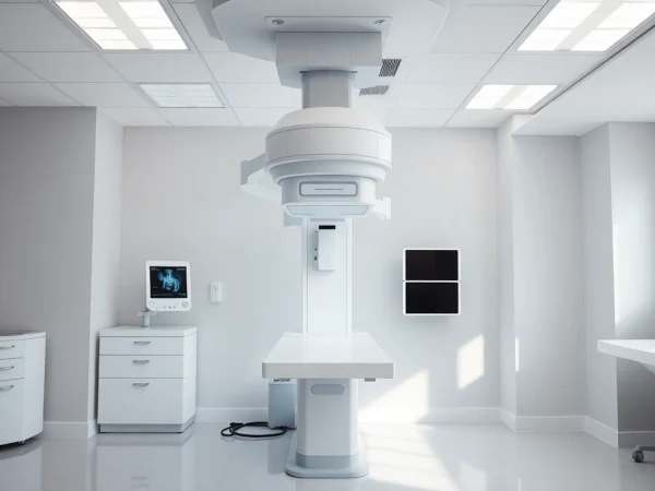
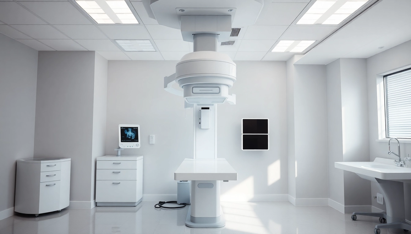
A veterinary x-ray system is a sophisticated imaging technology utilized in veterinary practices to diagnose and monitor a range of health conditions in animals. This equipment employs x-ray radiation to create images of the internal structures of an animal, aiding veterinarians in assessing skeletal injuries, tumors, organ anomalies, and other medical conditions. Unlike standard x-ray systems designed for humans, veterinary x-ray systems are specifically calibrated to address the anatomical and physiological characteristics of various animal species. This tailored approach ensures higher accuracy and patient safety while providing critical insights into a pet’s health.
How Veterinary X-Ray Systems Work
The functionality of a veterinary x-ray system revolves around the principles of x-ray radiation and image formation. When an animal is positioned properly in the machine, it is exposed to controlled doses of x-ray radiation. These rays penetrate the tissues and organs and are absorbed by different structures to varying degrees, resulting in a shadow-like image on a digital detector or film. The denser the structure—such as bones—the more radiation it absorbs, appearing white on the developed image. Conversely, less dense structures, like muscles and fat, allow more radiation to pass through, appearing darker. The transition to digital x-ray systems has revolutionized this process, allowing for immediate image review and the ability to enhance images for better diagnostics.
Benefits of Using X-Rays in Veterinary Medicine
Utilizing x-ray technology in veterinary medicine offers numerous advantages. Firstly, it provides invaluable insight into the health status of pets without the need for invasive procedures. Quick, efficient diagnostics can significantly reduce the time taken to make informed treatment decisions, ultimately benefiting the pet’s recovery process. Additionally, x-rays are instrumental for pre-surgical planning, helping veterinarians assess the condition before surgery.
Furthermore, the clarity of x-ray images enables a better understanding of various health conditions, including fractures, dental issues, and internal organ diseases. This increased clarity contributes to more accurate diagnoses, higher treatment success rates, and an overall enhancement in the quality of care pets receive. Importantly, with advancements in technology, modern veterinary x-ray systems are designed with safety features that minimize radiation exposure for both the animals and the veterinary staff.
Choosing the Right Veterinary X-Ray System
Key Features to Look for in a Veterinary X-Ray System
When selecting a veterinary x-ray system, there are several crucial features that practitioners should consider:
- Image Quality: High-resolution imaging capabilities ensure that abnormalities are identified accurately.
- Digital Capability: Digital x-ray systems offer rapid image processing and storage options, allowing for easy comparisons and documentation.
- Equipment Portability: In clinics with limited space, portable systems or those with compact designs can be a major advantage.
- Ease of Use: User-friendly interfaces can significantly enhance workflow efficiency and minimize technician training time.
- Safety Features: Incorporating advanced safety features like lead shielding for operators and adjustable radiation output is crucial.
- Integration with Practice Management Software: A system that connects easily with existing software will enhance operational efficiency.
Comparing Different Veterinary X-Ray Systems
There are various types of veterinary x-ray systems available on the market, from traditional analog systems to advanced digital radiography. When comparing options, it’s essential to consider not just the technological capabilities but also the manufacturer’s reputation, customer support, and warranty services. Brands that are well-regarded in the industry often provide not only reliable equipment but also comprehensive training and support, which are critical for successful implementation.
Moreover, clinics should assess the overall cost versus the expected return on investment. While upfront costs can be high for advanced digital systems, the efficiency, reduced film expense, and improved diagnostic abilities often lead to long-term financial benefits.
Cost Considerations for Veterinary Practices
The investment in a veterinary x-ray system can vary widely based on features, technology, and brand. New digital systems generally range from $30,000 to $100,000, while used or refurbished equipment may offer significant savings. However, the initial expenses are only part of the equation; ongoing operational costs—including maintenance, consumables, software updates, and training—should also be factored in.
Veterinarians should also evaluate financing options or leasing arrangements that may help alleviate the financial burden. Many veterinary suppliers offer flexible payment plans, enabling practices to obtain high-quality imaging systems without upfront costs that could strain finances.
Best Practices for Operating a Veterinary X-Ray System
Safety Measures for Pets and Technicians
Ensuring a safe operating environment is paramount when using veterinary x-ray systems. Both the animals and technicians must be protected from unnecessary radiation exposure. Key safety measures include:
- Physical Barriers: Use lead barriers and aprons to shield staff from radiation.
- Distance: Technicians should maintain a safe distance from the radiation source during exposure.
- Regular Equipment Checks: Conduct routine maintenance and safety checks to ensure the x-ray equipment is functioning correctly.
- Training: Comprehensive training for all staff members on x-ray protocols and safety measures is essential.
- Calm Animal Handling: Employ gentle handling techniques to minimize stress for pets, which can sometimes influence the accuracy of imaging.
Effective Techniques for Imaging
Correct positioning of the animal is critical for obtaining clear and useful x-ray images. Technicians should be familiar with the anatomy of various species to optimize positioning correctly. Additionally, the use of positioning aids, such as foam pads or sandbags, can help keep pets in the desired position while ensuring comfort. Furthermore, understanding the appropriate settings for exposure based on the size and type of animal being imaged is essential to reduce motion blur and improve clarity.
Making use of digital enhancements, such as image manipulation software, can help to improve contrast, highlighting areas that may be of concern or enhancing detail in soft tissue imaging.
Handling Emergencies During X-Ray Procedures
Veterinarians and technicians must be prepared to handle emergencies that may arise during x-ray procedures. This could include managing a distressed animal that may attempt to escape or a situation where the animal experiences sudden health complications—especially in cases of trauma where immediate imaging is critical.
Establishing a clear protocol for emergencies, including updated contact information for veterinary anesthesiologists or ER personnel, ensures that staff are prepared. Practicing situational drills can aid in ensuring that all team members are responsive and efficient during real emergencies, providing peace of mind amidst high-stress situations.
Interpreting X-Ray Results in Veterinary Medicine
Common Conditions Diagnosed with Veterinary X-Rays
Veterinary x-ray systems play a crucial role in diagnosing numerous conditions, including:
- Fractures: Identifying broken bones is one of the primary uses of x-rays in veterinary medicine.
- Arthritis: X-rays can reveal joint irregularities and signs of degeneration.
- Dental Problems: Dental radiography identifies issues beneath the gum line and bone loss linked with periodontal disease.
- Tumors: Internal tumors can often be located through imaging.
- Organ Enlargement: Conditions like heart disease can be diagnosed by observing the heart size on x-rays.
Understanding X-Ray Images
Reading x-ray images requires a base understanding of xenographic principles and experience. Veterinarians often consult specialized radiologists for complex cases. Features to note on x-ray images include:
- Bone Density: Areas that appear darker may indicate osteopenia or bone loss.
- Soft Tissue Contrast: Understanding variations in the shades of gray can aid in identifying masses or lesions.
- Anatomical Positioning: Recognizing the appropriate anatomical relationships among structures is critical to diagnosis.
In modern veterinary practices, integrating artificial intelligence (AI) software is emerging as a potent tool to aid in the interpretation of x-ray results, potentially flagging abnormalities that may be easily overlooked.
Consulting with Veterinary Specialists
Collaboration with veterinary specialists can enhance diagnostics. In cases requiring advanced expertise, such as orthopedic surgery or oncology, consultation ensures that the pet receives the most informed care. Many veterinary practices have established networks with specialists, allowing for referrals and shared insights, ultimately leading to improved outcomes for animals.
Fostering such professional relationships can also help practices stay updated on new protocols and emerging technologies, leveraging collective knowledge for enhanced patient care.
The Future of Veterinary X-Ray Technology
Innovations in Veterinary Imaging Systems
The landscape of veterinary imaging is continually evolving, with innovations enhancing the capabilities of x-ray systems. Advancements such as computed tomography (CT) and magnetic resonance imaging (MRI) are becoming more accessible for veterinary practices, providing detailed insights that pair well with traditional x-ray imaging. Moreover, the advent of portable imaging solutions allows veterinarians to conduct imaging sessions in various environments, from traditional clinic settings to mobile hospitals serving rural areas.
Additionally, the integration of machine learning and AI into imaging software is proving revolutionary, offering automated analysis and quicker diagnosis, which not only enhances efficiency but may increase diagnostic accuracy.
The Role of Digital Technology in Veterinary X-Rays
Digital x-ray technology has reshaped the veterinary imaging landscape. The rapid turnaround time for producing images has allowed veterinarians to offer immediate care, which can be crucial for a pet’s health. Digital systems also facilitate cloud storage options, enabling easy access to imaging histories and cross-referencing cases quickly. Furthermore, the ability to share images electronically with specialists around the globe fosters collaborative diagnostics.
On the economic front, digital x-rays decrease the costs associated with film-based systems, with less wastage and lower recurring expenses for materials. This shift towards digital formats underscores a trend toward increased efficiency and eco-friendliness in veterinary practices.
Emerging Trends in Veterinary Radiology
As technology continues to progress, several trends are emerging in the field of veterinary radiology:
- Telemedicine: The rise of telemedicine in veterinary practices enables remote consultations, simplifying access to specialist opinions on complex cases.
- Mobile Imaging Units: Veterinary practices are increasingly adopting mobile imaging services, allowing for on-site diagnostics in convenient locations for pet owners.
- Enhanced Training Tools: Advanced simulation programs for radiologists are being developed, enabling training on complex diagnostic techniques in a controlled environment.
- Increased Focus on Preventative Care: With better imaging technology, the emphasis is growing on using x-rays for preventative care, helping to identify issues before they escalate.
These trends highlight a clear trajectory towards more efficiency, accuracy, and proactive care in veterinary practices, ultimately improving health outcomes for companion animals.